
.jpeg)
Clinical Research
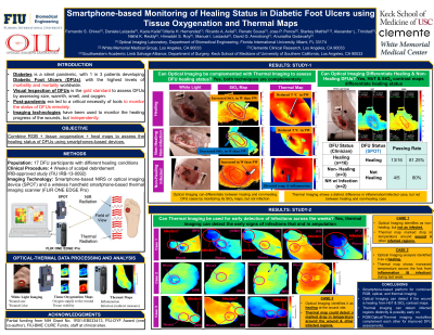
Fifteen DFU patients were recruited via a written consent form in this IRB approved study. Non-invasive and non-contact imaging measurements were performed using smartphone-based devices to measure for tissue oxygenation and thermal maps of the DFUs and its surroundings in each subject and across weeks of treatment. The tissue oxygenation maps in terms of oxygen saturation were used along with the thermal maps of the DFUs and its surrounding regions to determine the healing status of the DFUs (healing, non-healing, infectious, etc).
Results: Preliminary assessment of a healing and non-healing DFU case was performed across two weeks of the treatment process and the tissue oxygenation and thermal maps were compared. Both the patients were post-amputees with neuropathy and vascular issues. In the healing DFU case, the wound had increased temperature than its immediate surroundings by approximately 4 oC. On the contrary, this temperature difference increased to ~ 7 oC in a non-healing case. In terms of tissue oxygenation, the healing case showed a more homogeneous distribution within the wound and its immediate surroundings. On the contrary, it varied notably in the non-healing case.
Discussion: The thermal and tissue oxygenation maps tend to show varying distributions in a healing vs a non-healing DFU. The combined thermal and tissue oxygenation maps have the potential to differentiate healing from stalled healing (or non-healing) DFUs. Ongoing efforts are to determine quantitative metrics using combined optical-thermal maps across all 15 DFU cases to differentiate not only healing from non-healing (or stalled healing) DFUs, but also infectious and necrotic wounds.