(WHS-P36) Differences In Subcutaneous Thickness and Location Affecting Wound Healing in the Swine Model
Friday, May 17, 2024
7:30 AM - 5:00 PM East Coast USA Time
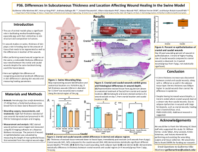
The use of the domesticated swine proves to be an integral part of advancing our understanding of dermatology in human skin due to its similarities to humans and the use of it as a model. A few reports in the literature have noted differences in healing in excisional wounds in different cephalo-caudal anatomical locations. We hypothesized that the differences in healing may be related to possible differences in the thickness of the dermal or adipose layers, as they contribute to the healing process. Here we addressed this question of how these layers vary in thickness when excisional wounds are created in different anatomical locations, albeit when excising down to the same anatomical landmark of the panniculosus carnosus muscle in the attempt to create equivalent wounds. Eight full-thickness wounds (1.6cm in diameter for 2cm.2 area) were created along the dorsal region of the in Yorkshire/Landrace crossbreed pigs along a cephalo-caudal axis, with each wound being 3cm separated from the adjacent one. Both the depth of the resultant wound and the excised tissues were measured. At day 7 post-wounding, the entire wound was excised and fixed and sectioned, and the thickness of the dermal and subdermal tissues at the wound margin was measured histomorphometrically. Wound depths of the excised cranial wounds averaged 8.8cm while those in the caudal area averaged 6.7cm (N=28 wounds, 0.01 P-value). Histomorphometric analysis of the dermal and subdermal layers adjacent to the excised wounds showed that in the cranially located wounds the dermis averaged 2515um as compared to 2747um in caudally located wounds (N=24, 0.05 P-value), and the subcutaneous layers measured 6674um and 4584 in the cranial and caudal wounds respectively (N=24, 0.0001 P-value). This study demonstrates that despite excising down to the same anatomical landmark in an attempt to create identical wounds, the dermal and subcutaneous tissues are thicker in cranial areas as compared to the caudal wounds. Surprisingly, however, on day 7 post wounding, there is no difference in the wound re-epithelization between these two areas. Additional studies examining healing at later time points may reveal differences in healing.

.jpeg)