(WHS-P30) NOVEL PEEL AND PLACE NEGATIVE PRESSURE WOUND THERAPY DRESSING PRECLINICAL EVALUATION: FINITE ELEMENT MODELING OF WOUND BED TISSUE STRAINS EXPANDS UPON BIOMARKER OUTCOMES
Friday, May 17, 2024
7:30 AM - 5:00 PM East Coast USA Time
Purpose: A novel peel and place† dressing for use with negative pressure wound therapy (NPWT)* has been developed that addresses the challenges of tissue ingrowth with reticulated open cell foam (ROCF)^ dressings, while exhibiting a modified tissue strain environment and promotion of wound healing associated biomarkers. Materials and
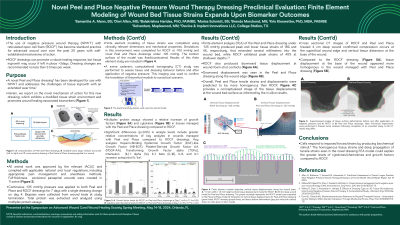
Methods: All animal work was approved by the relevant IACUC and complied with applicable national and local regulations. Full-thickness excisional paraspinal wounds were created in 11 swine. Continuous, -125 mmHg was applied to both peel and place and ROCF dressings for 7 days with a single dressing change on day 4. Biopsies were collected from wound beds at study termination. Total protein was extracted and analyzed using multiplex protein assays. Finite element modeling of tissue strains was completed using clinically relevant dimensions and mechanical properties. Simulations in this environment were completed for ROCF and novel peel and place NPWT dressings under -125 mmHg. Results/
Discussion: Multiplex protein assays showed a relative increase of cytokines and growth factors in tissues managed with the peel and place dressing compared to ROCF. Notable significant differences (p≤0.05) include greater relative concentrations of HB-EGF (19.36 vs. 5.381 ng/g), PDGF-AA (15.46 vs 11.14 ng/g), TGF-a (8.253 vs. 1.861 ng/g) in peel and place managed wounds compared to ROCF. Finite element analysis (FEA) of novel dressing under -125 mmHg produced peak and lower tissue strains of 18% and 4%, respectively, that extended several mm into the wound bed, while ROCF exhibited peak strains of 40% at shallower depths. ROCF also produced downward tissue displacement at wound-foam strut contacts. Downward displacement was seen in the peel and place dressing along the wound edge. Overall, tensile strains were predicted to be more homogenous at the deep wound bed in the peel and place dressing model.
Conclusion: Cells respond to imposed forces/strains by producing biochemical stimuli.∏ The homogenous tissue strains and deep propagation of tensile strains seen in the novel dressing FEA model could explain the greater levels of cytokines/chemokines and growth factors compared to ROCF. †Novel Peel and Place Dressing (3M Company, San Antonio, TX); * 3M™ V.A.C.® Therapy; ^ 3M™ V.A.C.® Granufoam™ ∏ Dunn, S. L., & Olmedo, M. L. (2016). Mechanotransduction: Relevance to Physical Therapist Practice Understanding Our Ability to Affect Genetic Expression Through Mechanical Forces. Physical Therapy, 96(5), 712-721. doi:10.2522/ptj.20150073

.jpeg)