(CR-015) Differentiate Healing, Non-Healing, and Infectious Diabetic Foot Ulcers Using a Smartphone-based Thermal Scanner
Thursday, May 16, 2024
7:30 PM - 8:30 PM East Coast USA Time
Fernando Chiwo, Daniela Leizaola, Ricardo A. Avila, Renato Sousa, Jose P. Ponce, Stanley Mathis, David G. Armstrong, Anuradha Godavarty
Introduction: Diabetic foot ulcers (DFUs) have become a global health project in which many efforts have been discussed for early detection and prevention. Thermal imaging has become a promising tool for DFUs imaging due to its non-invasive and non-contact measurement that is available as a hand-held imaging tool to objective assess healing status of DFUs.In this study, a smartphone-based thermal scanner was employed to differentiate healing, non-healing, and infectious DFUs.
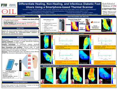
Methods: Seventeen DFU participants with different healing conditions of DFUs were imaged across six weeks of their weekly treatment after a scalpel debridement process. A commercial handheld smartphone-based thermal imaging scanner was used to image the wound and periwound surfaces across the foot area. Thermal maps were used to classify if the DFUs were healing, stalled healing (or non-healing), and/or infected. The thermal index was calculated as the ratio of the temperatures between the wound and periwound at selected point locations.
Results: From a preliminary analysis of the thermal maps at point locations in the wound and peri-wound, the following was observed: (i) the healing DFU case showed a homogeneous thermal distribution in the overall foot, with a slightly increased temperature on the wound surface; (ii) the stalled healing (or non-healing) DFU cases showed a higher variation in temperature of the full foot, also with a contrasted difference in temperature between the wound and periwound; and (iii) the infected DFU case showed the highest temperature increase of the three cases within the wound region. However, the remaining foot area showed a similar distribution to the healing cases.
Discussion: A smartphone-based thermal imaging device has the potential to differentiate healing, non-healing, and infected DFUs based on the temperature distributions of the foot.Our ongoing studies are focused on developing quantitative thermal indices using data obtained across all the DFU cases and across the weeks to develop a thermal indicator of the wound status in DFUs.This can assist in determining onset of infections, potential to heal, and/or the underlying necrosis in these tissues.

.jpeg)