
.jpeg)
Laboratory Research
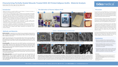
Personalized grafts, tailored to the individual, can now be created using a groundbreaking 3D printing process that uses the patient's own adipose tissue as bioink. This process involves homogenizing liposuction-derived aspirate into a bioink that is rich in autologous growth factors and cells, making it conducive to healing. This bioink was used in an FDA-cleared extrusion-based 3D bioprinter. These innovative grafts aid in the healing process and act as scaffolding, promoting granulation and re-epithelialization in wound beds.
Control of certain scaffold characteristics such as mechanical properties, morphology, and nanoscale porosity is key to optimizing tissue regeneration and remodeling. This study characterizes the porosity and morphology of the 3D printed adipose scaffolds using scanning electron microscopy (SEM), and assesses their mechanobiological properties, including elasticity, surface roughness, and adipocyte interaction with the scaffold, through nanoindentation. Setting target ranges for these critical material properties of the adipose wound grafts is important in establishing a standard for skin tissue regeneration.
Methods:
Rat Model:
Full-thickness defects in 450 g Sprague Dawley rats were used to assess the healing of 3D printed human adipose grafts. Four 8 mm defects were created dorsally, and filled with 1 cm samples. Healed defects were explanted on days 7 and 14.
Adipose 3D Printed Graft:
The adipose tissue aspirate was homogenized and washed in saline. A novel FDA-cleared 3D printed was used to create the 3mm thick wound grafts. Grafts were solidified via temperature control before implantation.
Scanning Electron Microscopy:
A Zeiss Gemini 360 FE-SEM imaged the tissue samples.
Nanoindentation:
Using a Pavone Optics 11 Life nanoindenter, the elasticity and surface roughness of explanted samples was measured in a 25 μm x 25 μm area. This was followed by light microscopic analysis. Samples were soaked in Phosphate Buffer Saline (PBS) for 24 hours and affixed to the surface with cyanoacrylate to prevent movement during testing.
Results:
The study presents SEM images of the adipose wound graft, including pore size analysis. Additionally, the elasticity of the graft was quantified across a 25 μm x 25 μm area.
Discussion: