(CR-004) The Use of Near-Infrared Spectroscopy in Assessment of Viability and Monitoring of Healing Trajectory in Traumatic Flaps
Thursday, May 16, 2024
7:30 PM - 8:30 PM East Coast USA Time
Homer-Christian Reiter, BSc
Introduction: Patients are frequently treated in wound care for traumatic flaps. Often these acute wounds are covered by anatomically displaced skin which is removed by providers despite its ability to act as a skin graft. If flap tissue is still adequately perfused and there is no infection, the base of the wound should be cleaned, and the flap should be tacked in proper anatomical position with steri strips. This benefits the patient by protecting the wound with epidermal tissue that can adhere to intact dermis in lieu of creating an uncovered wound that must re-epithelialize. Before this tissue may remain, however, it must be assessed for viability. A necrosed flap introduces an infection risk to the wound bed and may delay healing, so it is essential to monitor the vitality of flap tissue each subsequent visit. This can be done primarily by clinical assessment, but visual inspection does not offer the precision of near-infrared spectroscopy (NIRS) which can give information on oxygenation and hemoglobin content of the tissue.
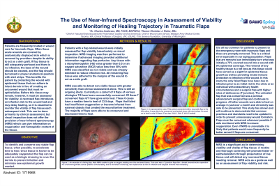
Methods: Patients with a flap related wound were initially assessed for flap viability based solely on visual inspection. NIRS imaging was then performed to determine if advanced imaging provided additional information regarding flap perfusion. Any tissue with a deoxyhemoglobin (Hb) value greater than 0.5 or an oxygen saturation (StO2) value less than 50% with NIRS was classified as non-viable and was sharply debrided to reduce infection risk. All remaining flap tissue was adhered to the margins of the wound to act as a skin graft.
Results: NIRS was able to detect non-viable tissue with greater sensitivity than clinical assessment alone. Flaps that failed had insufficient oxygenation or became infected from external objects that created the wound before treatment. The majority of flaps were able to be conserved and decreased time to heal.
Discussion: NIRS is a significant aid in determining viability and vitality of flap tissue. A routine and thorough screening will promote faster re-epithelialization through preservation of live tissue and will detect any necrosed tissue needing removal. NIRS acts as a guide as well as an assessment of flap viability and risk.

.jpeg)