(LR-021) Wireless Closed-Loop Biomimetic Nanoplasmonic Dressings for Spatiotemporal Imaging and Therapy of Pseudomonas Aeruginosa Biofilms in Wounds
Thursday, May 16, 2024
7:30 PM - 8:30 PM East Coast USA Time
Meitong Nie, PhD; Stacie Deaver, PhD; Benjamin Pittelkau, BS; Ze Zong, BS; Erin Gloag, PhD; Wei Zhou, PhD
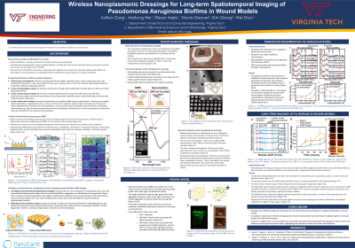
Introduction: Recent studies indicate that early and continuous detection and eradication of heterogeneously distributed pathogenic biofilms in wounds significantly enhance wound healing outcomes.1 Despite advancements in wearable diagnostics, the continuous large-area imaging of infection markers without removing bandages remains a significant challenge. This challenge stems from several factors, including the limited spatial resolution of conventional individually-addressable electrode-based electrical sensors, sensor contamination due to the complex composition of wound media, and the necessity of bandage removal for standard optical imaging techniques.
Methods: We have developed biomimetic nanoplasmonic dressings—a fusion of multiresonant nanoplasmonic elements on a biomimetic polymeric scaffold.2,3 This design facilitates inflammation-free sensing and actuation directly at the wound bed. The multiresonant nanoplasmonic elements enable ultra-sensitive, label-free molecular imaging of biofilm components through Surface-enhanced Raman Spectroscopy (SERS) with exceptional spatial resolution (1 μm) across extensive areas (10 cm2).2,3 For continuous monitoring, our technology ensures sensor surface regeneration via pulsed-laser-induced plasmonic nanocavitation. Leveraging machine learning on SERS datasets, we analyze dynamic spatiotemporal biofilm evolution and identify pathogenic bacteria within in-vitro wound models.3,4 Our agar block biofilm assay for in-vitro wound modeling utilizes P. aeruginosa inoculated into wound-like media with agar, mimicking biofilm growth in-vivo.5 Furthermore, our research extends to closed-loop therapy of biofilms, employing a synergistic combination of antibiotics and plasmonic photothermal-photoacoustic effects.
Results: Our nanoplasmonic devices exhibit outstanding SERS enhancement factors ( >107) with a minimal relative standard deviation of 11.5%.3 We successfully detect pyocyanin, a P. aeruginosa virulence factor, at concentrations as low as 10-8M in a blood serum background after a 24-hour device incubation. This sensitivity is crucial as typical pyocyanin concentrations in infected wounds at early stages range from 0.1-100 μM.6 Additionally, we demonstrate large-area spatiotemporal imaging of pyocyanin secretion by P. aeruginosa within in-vitro wound models over 32 hours, revealing unique spatiotemporal evolutions across various mutant strains. Pyocyanin signals are detected within 6 hours of culturing, indicating the potential for early detection. Finally, a substantial reduction in viable bacteria is observed after combination treatment, validated through cfu analysis and live-dead imaging.
Discussion: Our technology shows promise for ultrasensitive (< 10-8 M pyocyanin), large-area ( >10 cm2), and prolonged ( >32 hours) imaging of P. aeruginosa, coupled with on-demand therapeutics. This wireless system integrates miniaturized optical instrumentation with nanoplasmonic dressings through user-friendly software, offering the prospect of convenient wound biofilm management without bandage removal. However, studies in animal models and with diverse bacterial strains are essential to validate the technology's clinical potential.

.jpeg)