Case Series/Study
(CS-056) Ultra-Thick Amniotic Membrane Allograft and Disposable Negative Pressure Wound Therapy in the Management of an Ischemic, Diabetic Ulcer
Thursday, May 16, 2024
7:30 PM - 8:30 PM East Coast USA Time

Introduction: Lower extremity ulceration is an increasingly common and debilitating condition, affecting 1-2% of the world’s population and up to 5% of the elderly population.1 P</span>eripheral vascular disease is a dominating causative factor of these ulcers which can be especially difficult to heal in patients with multiple risk factors such as Diabetes, ischemia, and history of smoking.2, 3 To aid the healing in such complex cases, adjunctive treatment measures may be warranted such as negative pressure wound therapy (NPWT) and Amniotic Membrane (AM) to help address the physiological deficiencies underlying the wound.4-7
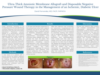
Methods: A case report of a patient with a wound around the lateral malleolus complicated by multiple risk factors for healing that was successfully treated with cryopreserved, ultra-thick amniotic membrane (AM) allograft† derived from umbilical cord and disposable NPWT+.
Results: A 57-year-old female presented to the wound care clinic with a lateral malleolar wound on the right foot. The patient had a medical history of Diabetes, peripheral vascular disease (PVD), hypercholesterolemia, hypertension, neuropathy, and coronary artery disease and was a smoker for over 40 years. The patient received multiple prior treatments including wound debridements with hydrocolloids, right axillary and femoral popliteal bypass surgery for PVD, compression dressings for venous insufficiency, and systemic and topical antibiotics for multidrug-resistant Achromobacter-infection of the wound. After the infection was controlled, AM and disposable NPWT was placed on the 2x 1x 0.2cm wound to promote healing and reduce risk of underlying tissue exposure. Increased blood perfusion and epithelialization were noted over the next five weeks and the wound decreased in size to 1.4x 0.8x 0.2cm. Repeat AM and NPWT application were performed at five and six weeks. The wound continued to epithelialize and completely heal at nine weeks after the initial AM and NPWT treatment.
Discussion:
This case highlights the unique application of ultra-thick AM along with disposable NPWT to support wound closure in a wound complicated by multiple risk factors.

.jpeg)